Revolutionizing Chest X-Ray: A New Neural Network Model

Scientists have achieved a significant breakthrough in a pivotal report recently published in Radiology about New Neural Network Model. By joining clinical patient information with imaging information, they had the option to work on the analytic viability of chest X-beams essentially. The field of clinical imaging could go through an unrest because of this disclosure, which would likewise significantly improve patient consideration.

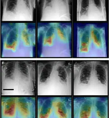
The presentation includes anteroposterior radiographs (top) and corresponding attention maps (bottom) taken with patients in a supine position.

A. The top row of images showcases the most critical diagnostic findings from our proprietary dataset. A 69-year-old female patient with blockage, pneumonic invades, and emission is shown on the left. In the center, a 64-year-old with similar symptoms, and on the right, a 49-year-old with cardiomegaly and pneumonic infiltrates.

B. The bottom row highlights significant diagnostic findings from the Medical Information Mart for Intensive Care dataset. A 48-year-old female patient with pneumonic penetrates in the lower right lung is shown in the center. On the left, a 58-year-old female patient with respective atelectasis and lower lung emanation. On the right, a 79-year-old male patient with cardiomegaly and pneumonic penetrates in the right lower lung were the three cases that were introduce

New Neural Network Model image
Chest X-Ray Diagnostic-New Neural Network Model

It’s essential to note that our attention maps consistently identify the most relevant regions within the images. For instance, when pneumonic opacities are present, these maps clearly indicate those areas in the lungs. Credit for these images goes to the Radiological Society of North America.

When healthcare professionals diagnose illnesses, they use both imaging and non-imaging data. Current AI-based methods often work with data separately to solve tasks.
 
Transformer-based brain organizations, an arising sort of man-made intelligence model, can possibly give more exact judgments by joining imaging and non-imaging information. These Transformer models were initially made for normal language handling undertakings with the assistance of PCs. They have now tracked down applications in engaging huge language models like ChatGPT and Google’s man-made intelligence visit administration Troubadour, supporting their abilities in dealing with complex language assignments.
 

Processing Data 

Unlike convolutional neural networks, designed for processing imaging data, Transformer models adopt a broader neural network architecture. They depend on a consideration component, which permits the brain organization to learn connections between components in its feedback. 
 
Toward the day’s end, this component is particularly useful in the clinical region, where various components like patient information and imaging results much of the time assume critical parts in determination.
 
Faraz Khadar, Study Lead Creator and PhD Competitor in the Branch of Symptomatic and Interventional Radiology at College Clinic Aachen.
This capacity holds extraordinary commitment in the field of medication, where different variables, including patient information and imaging results, much of the time contribute essentially to the symptomatic cycle.
 

Model Trained on Imaging and Non-Imaging Data from 82K Patients

Khadar and his colleagues specifically developed a Transformer model for medical applications. They trained it on both imaging and non-imaging patient data, including information from over 82,000 patients.

 This model was trained to detect up to 25 different anomalies, whether using non-imaging data, imaging data, or a combination of both, referred to as multi-modal data.
 
The multi-modal model outperformed other models in terms of diagnostic performance for all cases, showing promise in aiding healthcare professionals in the future.
 
In the era of increasing workload, it holds the potential to assist healthcare providers effectively.
 
Khadar further explained, “With the continuous growth in the volume of patient data over the years, which can be daunting for doctors, as they have limited time per patient, analyzing all available information effectively can be challenging. Multi-modal models promise to simplify the aggregation of data, making it easier to aid healthcare providers in accurate diagnosis.”
 
In essence, the approved design can effectively correlate a significant amount of data.


Journal Reference:

Khader, F., et al. (2023) Multimodal Deep Learning for Integrating Chest Radiographs and Clinical Parameters: A Case for Transformers. Radiology. doi:10.1148/radiol.230806.

Source: https://www.rsna.org/

Source link